Ultrasound Examination in Pets
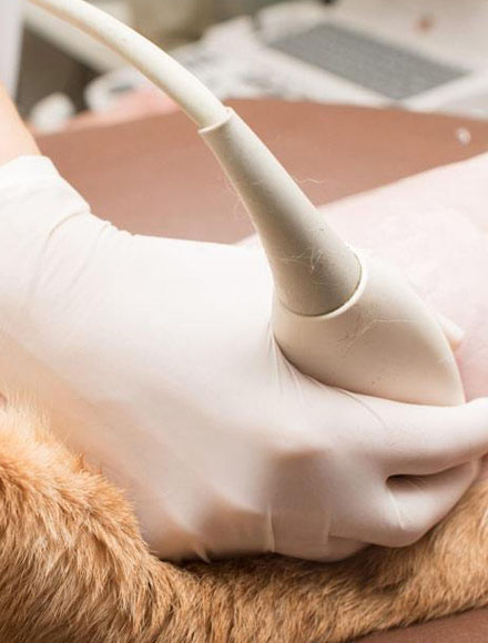
Ultrasound equipment directs a narrow beam of high frequency sound waves into the area of interest. The sound waves may be transmitted through, reflected, or absorbed by the tissues that they encounter.
The ultrasound is an imaging procedure that uses sound waves that aren’t audible to the human ear. These sounds “echo” off the corresponding site in question, which produces images that are mapped by black (fluid) and grey (tissue). From these images, we can get a better understanding of organ health and detect things like a tumor or pregnancy.
Is the technique affordable?
Although the initial cost of a scan may seem high, it has to be equated with the high cost of the equipment, the fact that specialized training is required in order to interpret the images, and a significant amount of time is involved in carrying out the examination. Its usefulness for pregnancy diagnosis, evaluation of the internal organs, assessment of heart function, and evaluation of certain eye diseases, make it an invaluable, non-invasive diagnostic tool to help protect to your pet’s well-being.
IS ULTRASOUND COMFORTABLE?
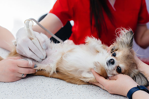
An ultrasound examination, also known as ultrasonography, is a non-invasive imaging technique that allows internal body structures to be seen by recording echoes or reflections of ultrasonic waves. Ultrasound is a non-invasive imaging technique that is considered safe and does not involve radiation.
Ultrasound equipment directs a narrow beam of high frequency sound waves into the area of interest. The sound waves may be transmitted through, reflected, or absorbed by the tissues that they encounter.
HOW DOES ULTRASOUND WORK?
The ultrasound waves that are reflected will return as echoes to the probe and are converted into an image that is displayed on the monitor, giving a 2-dimensional “picture” of the tissues under examination.
The technique is invaluable for the examination of internal organs and was first used in veterinary medicine for pregnancy diagnosis. However, the technique is also extremely useful in evaluating heart conditions and identifying changes in abdominal organs. Ultrasonography is very useful in the identification of cysts and tumors.
What Our Customers Are Saying
Come See Us!
If you live in the surrounding area and need a trusted veterinarian to care for your pets – look no further. Dr. Anshul Jindal is a licensed ON veterinarian, treating all types of pets. Your pets’ health and well being are very important to us, and we take every possible measure to give your animals the care they deserve.


